Unsere Schwerpunkte: Volkserkrankungen wie Asthma, Allergien und Chronisch Obstruktive Lungenerkrankungen (COPD), sowie infektionsbedingte Entzündungen der Lunge, vor allem die Tuberkulose (TB).

17.02.2026
Auch im Jahr 2026 ist Prof. Uta Jappe, Leiterin der Forschungsgruppe "Klinische und Molekulare Allergologie" am FZB wieder in der renommierten STERN‑Ärzteliste im Bereich Nahrungsmittelunverträglichkeiten vertreten. Die STERN‑Ärzteliste gilt als eines der umfangreichsten und bekanntesten Orientierungssysteme für Patientinnen und Patienten in Deutschland.

04.02.2026
Prof. Christoph Lange, Medizinischer Direktor des Forschungszentrums Borstel, Leibniz Lungenzentrum, erhält den Oskar-Medizinpreis 2025 der Stiftung Oskar-Helene-Heim. Der mit 50.000 Euro dotierte Preis zählt zu den höchstdotierten Medizinpreisen in Deutschland.

02.02.2026
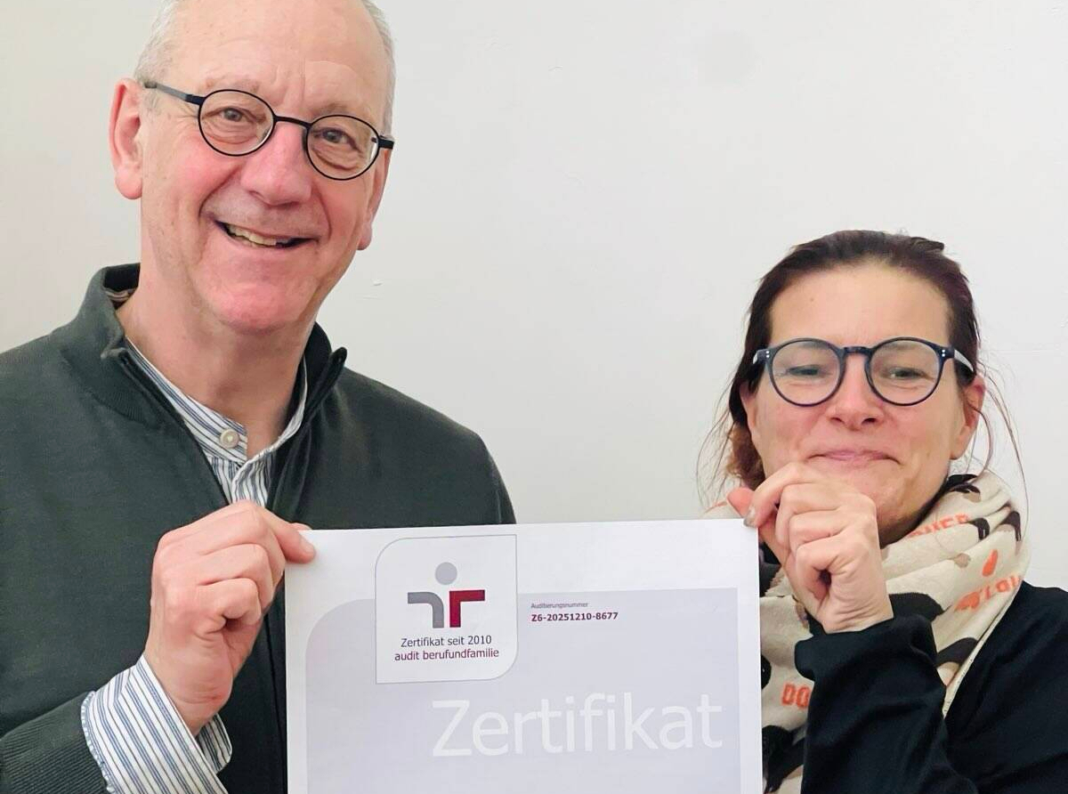
Das FZB hat vom Kuratorium der berufundfamilie Service GmbH die Bestätigung für das Zertifikat „audit berufundfamilie” erhalten. Damit sichert sich das FZB zum dritten Mal das Zertifikat mit Prädikat als besondere Anerkennung für seine langjährige, nachhaltige familien- und lebensphasenbewusste Personalpolitik. Das Zertifikat gilt als Qualitätssiegel für eine strategisch angelegte Vereinbarkeitspolitik.